
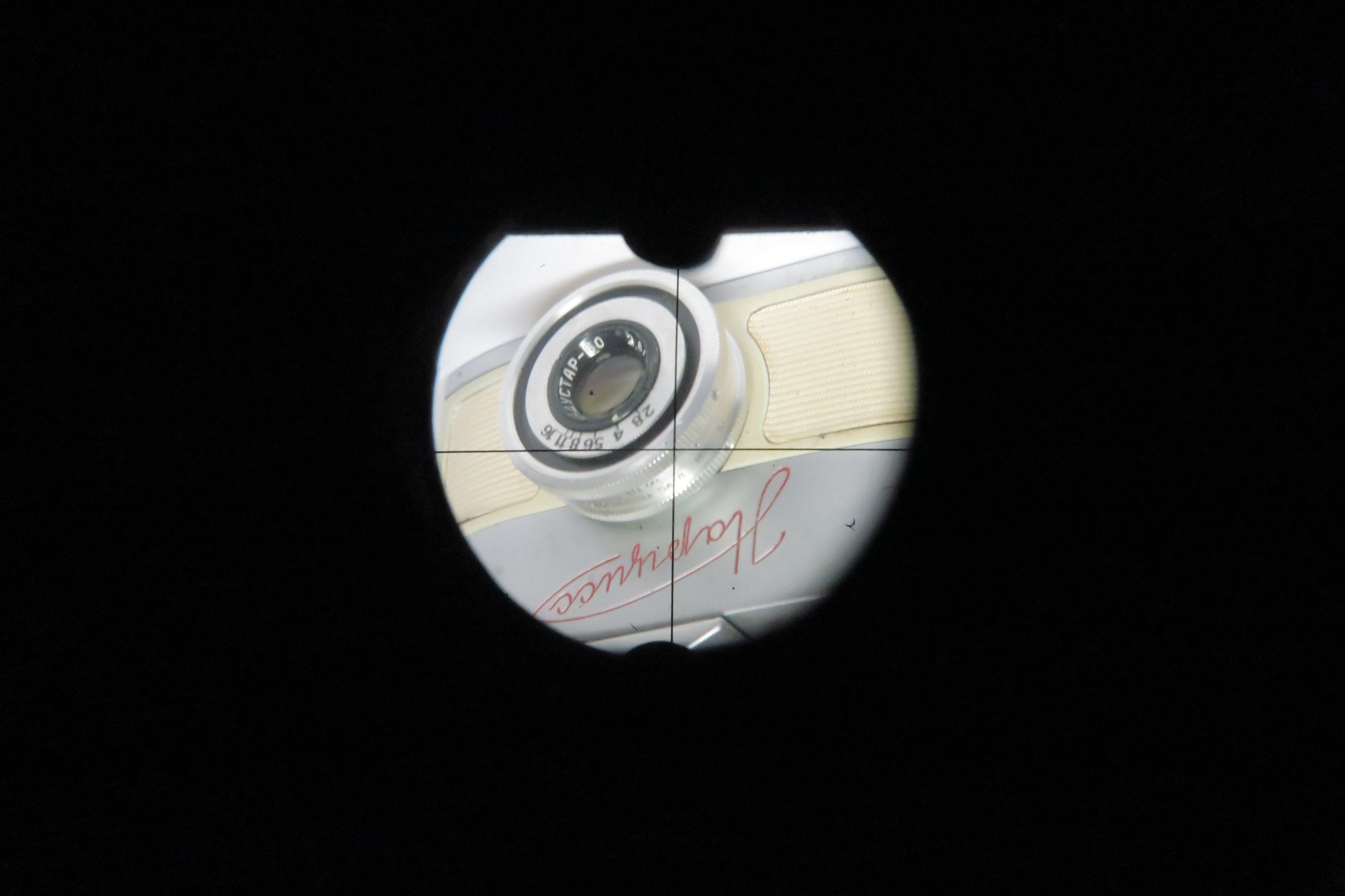
"By the way: one of the images I sent you probably was lost: the view through the endoscopic version of the „prism“ (which in this case is a simple mirror rather). It shows that the viewfinder image is indeed round and bottom-up (and unreversed laterally, as can be seen from the engraving „Narciss“).
Also by the way: when I was in the hospital, in the 1970/80s, my colleagues did many endoscopies using an Olympus endoscope (at the time the best available), and the Olympus Pen F SLR camera (half-frame 18x24 on film) was used for documentation. It is funny that the camera they used also was a special version, namely a special version of the Pen F, with round viewfinder image and cross-hair, no ground-glass, but a simple clear glass. I do not think that one firm (KMZ and Olympus) copied from the other, I believe that the purpose simply demands for a certain tool, but – it looks like that the Narciss (dating from the early 1960s) was ahead of the Olympus Pen F (introduced 1963 in the plain form, and in the endoscopic form probably some years later). Anyway, I believe that the form of the special version of the cameras simply came from the need to meet the requirements.
Another time (3rd time) by the way: there also was a discussion whether the small format of the Narciss was a drawback when the camera was used for such important things as endoscopic examinations. But the question is irrelevant in reality. Diagnoses in gastroscopy or coloscopy are never made by simply looking at (or photographing) a detail. Of course the examiner in most cases has a strong feeling as for „harmless“ or „bad/malignant“ when he sees a detail, but: final diagnoses are made by means of a biopsy exclusively. You cut a little specimen off the region of interest and the examination of this in a microscope will tell you the truth (which will take a little time, as the specimen must be soaked with a special paraffin or plastics or deep frozen, then cut into thin slices and stained with special dies). The photographs that are usually taken during an endoscopy are a sort of a reminder of the visual impression you got during endoscopy, and they are mostly combined with the verbal description of what was seen through the endoscope in the endoscopy report (that is then delivered to the patient and his doctor), but the photographs never are the exclusive or decisive source of the final diagnosis. So the quality of these photographs is not as important as one might think. Very probably the advantage of a large depth-of-field was bigger than the disadvantage of lesser resolution. As I said: histology (the examination of the endoscopically taken specimen with the microscope) is the all-decisive tool in endoscopy."
Best regards,
Milos and Vlad.